Eric L. Crowell, MD, MPH and Amde Selassie Shifera, MD, PhD
A 62-year-old man presented with a 5-day history of central scotoma of his left eye. Five days prior to presentation he hit his head against the bottom of a trailer. Approximately 30 minutes after the accident he noticed a dark grayish spot in the center of his vision. He also reported that when looks at a light source the light would look reddish. The patient also reported having a generalized headache of a moderate degree that lasted for 15 minutes after the accident.
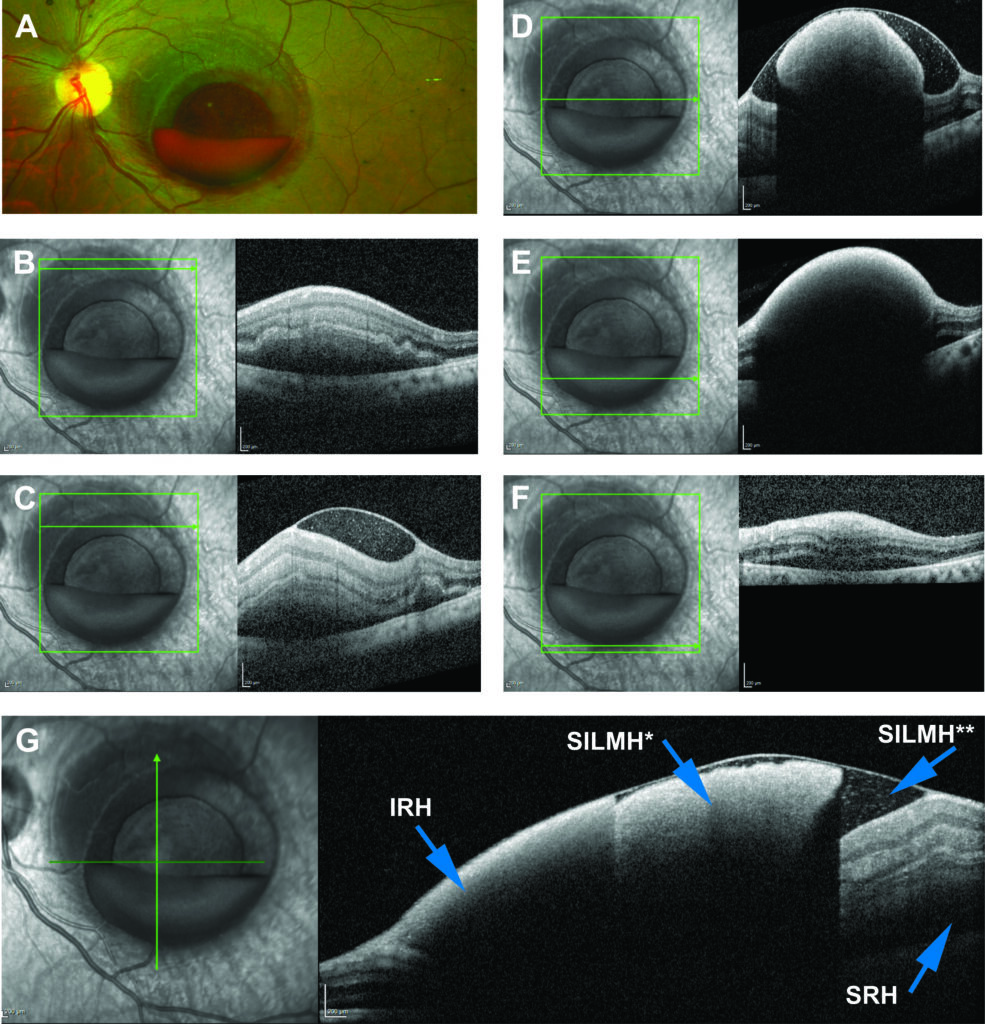
At presentation, his visual acuity in the left eye was counting fingers at 3 feet. On examination he was found to have a layered hemorrhage in the macula of the left eye (Figure 1A). Spectral domain optical coherence tomography (OCT; Spectralis system; Heidelberg Engineering) showed multi-layered hemorrhage in the macula of the left eye. Specifically, the OCT showed sub-internal limiting membrane (ILM) hemorrhage (both clotted and non-clotted), intraretinal hemorrhage and subretinal hemorrhage (Figure 1B to G). Examination of the right eye was non-remarkable. A computed tomography of the head done at presentation did not show any evidence of intracranial hemorrhage. No intervention was done at the initial presentation.


REFERENCES
1. Augsten R, Konigsdorffer E and Strobel J. Surgical approach in terson syndrome: vitreous and retinal findings. Eur J Ophthalmol 2000; 10:293-296.
2. Biousse V et al. The ophthalmology of intracranial vascular abnormalities. Am J Ophthalmol 1998; 125:527-544.
3. Gress DR, Wintermark M and Gean AD. A case of Terson syndrome and its mechanism of bleeding. J Neuroradiol 2013; 40:312-314.
4. Kapoor S. Terson syndrome: an often overlooked complication of subarachnoid hemorrhage. World Neurosurg 2014; 81:e4.
Eric L. Crowell, MD, MPH; Wilmer Eye Institute, Johns Hopkins University School of Medicine, Baltimore, Maryland (former address)
Amde Selassie Shifera, MD, PhD; Wilmer Eye Institute, Johns Hopkins University School of Medicine, Baltimore, Maryland (former address)
